The Cellular and Molecular Imaging (CMI) core facility of the Integrative Neuroscience COBRE center provides users with instruments and services for analyzing and quantification of biomolecules, and microscopic investigation of cells, tissues, organs, and small animals with imaging capabilities for brightfield and fluorescent microscopy. The core maintains several state-of-the-art microscopy instruments including a Leica SP8 confocal with LIGHTNING capabilities, a Scientifica two-photon microscope, a Leica THUNDER 3D-tissue and a Model organism with computational-clearing capabilities. The core also maintains multiple instruments for imaging, analyzing and quantifying DNA, RNA and protein such as a Typhoon Phosphoimager, automated PCR-pipetting robot and Real-Time PCR machines. Finally, the core has modern work space and storage for the culture of insect and mammalian cells with instruments including a Microfluidic Cell Sorter and microscope systems.
We like to keep track of our scientific contributions; please send us an email when your research is published. For grant proposals, we can provide support letters and core instrument information used for your research project. To acknowledge our core funding in your publications, please use P30 GM145646 and/or P20 GM103650. Use P20 for grant years from before July 2022 and P30 after; funding that spans both terms should use both numbers. Liquid Nitrogen Storage Tank
Location: FA 312 (door code access required)
Two Biorad real-time PCR machines combine advanced optical technology with precise thermal control to deliver sensitive, reliable detection, quick set-up runs and monitoring of amplification traces in real-time on the integrated LCD touch screen. Directly adjacent to the two qPCR detection systems, there is a PC computer containing easy to use software for on-site analysis of qPCR data.
Location: FA 312 (door code access required)
The Biorad Chemidoc is an easy, high-performance gel and blot imaging for accurate quantification of protein samples in gels and membranes. The Chemidoc streamlines Western blotting in a five-step approach by combining traditional blotting techniques with innovative tools to allow improved band quantitation and quick assessment of electrophoresis results and blot transfer quality prior to western blotting. The Chemidoc enables rapid western blot results in as little as 15 min with efficient protein transfer in 3-minute and 5-minute stain-free gel imaging. The embedded software includes all the tools for quantitative and qualitative image and data analysis. Images taken on the ChemiDoc can also directly be printed.
Location: FA 314 (door code access required)
The CellDrop FL is an automated cell counter that features a DirectPipette technology to eliminate plastic slides and cumbersome hemocytometers from routine cell counting. The CellDrop FL comes with dual channel fluorescence and brightfield optics, variable height sample chamber, and easy-to-use analysis software. It enables fast cell counts, viability assessments, and GFP transfection efficienty measurements across the widest range of cell density, cell type and application.
Location: FA 314 (door code access required)
The EVOS M5000 imaging system from ThermoFisher Scientific is a fully integrated digital inverted microscope for fluorescence, color and transmitted light for tissue culture dishes and post-cell sorting applications. It includes 3 filter cubes for imaging DAPI, Texas Red and GFP-labeled cells, and a high-resolution CMOS camera to capture quality color images of cells post-sort. In essence, the microscope would allow high-quality images and a rapid assessment of fluorescently labeled cells post-sort.
Location: FA 312 (door code access required)
The Typhoon Phosphoimager is a fast and versatile laser scanner for sensitive and quantitative measurements of radio-isotropic-labeled samples. The Typhoon is a non-confocal variable mode laser scanner offering high-speed and resolution for precise quantitation of proteins, nucleic acids and other biomolecules. The scanner comprises "filmless" autoradiography with storage phosphor screens. The laser can generate 16-bit images at up to 25 μm pixel resolution to give precise quantitation in gels and blots, with a high speed to scan gels of up to 240 x 250 mm in less than 2 min. Two phosphor screens with a size of 200 X 250 mm are available for autoradiography. The scanner can detect a broad range of isotopes in radiolabeled samples. Directly adjacent to the phosphoimager system, there is a PC computer with a software package that allows on-site quantitative and qualitative analysis of images and data.
The Integra media autoclave allows the rapid and gentle preparation of a 1-10 liter culture medium. Precise control of temperature, time and pressure during the sterilization process guarantees constant high quality. The mediaclave can be used for the preparation and sterilization of numerous types of culture media, including media for C. elegans research. The wide adding port facilitates the safe addition of supplements prior to dispensing. The mediaclave is connected to a peristaltic pump housed in a vertical flow workstation to fill Petri dishes and vials.
Location: FA 324 (door code access required)
The Leica TCS SP8 LIGHTNING confocal allows image information extraction with super-resolution acquisition that exploits the sub-diffraction lateral resolution of confocal microscopy. It allows to capture cellular details and observe dynamics with a resolution down to 120 nm, which is close to STED super-resolution microscope capabilities. The upright confocal is capable of simultaneous collection from up to 3 detection channels (two PMTs, one HyD) using multiple laser lines (diode 405, blue 488, green 552, red 638 nm). The confocal is configured with a conventional tandem scanner (8 kHz), a super Z-galvo stage type S, multiple objectives (10x, 20x, 25x, 40x, 63x), and stands on an anti-vibration table. The confocal is connected to a computer workstation equipped with Leica LAS-X software and a 30-inch monitor.
THUNDER imagers are a brand-new class of computational clearing high-resolution wide-field microscopes developed exclusively by Leica Microsystems. These imagers can acquire sharp data and high-quality images by instant removal of out-of-focus blur or haze inherent to thick 3D samples that are generally acquired using widefield microscopy. These imagers also significantly shorten the imaging workflow from image acquisition to data analysis with the new LAS-X navigator software. The THUNDER Imager 3D Tissue is a motorized upright microscope and is outfitted with an ultra-fast DFC 9000 GT/C camera, and is ideal for cell culture, tissues, fixed samples, and cleared tissues. The THUNDER Imager Model Organism is an M205 FCA stereomicroscope intended to image embryos, whole organs and small organisms.
Key features of the THUNDER 3D Tissue imager include:
Key features of the THUNDER Model Organism imager include:
Location: FA 312 (door code access required)
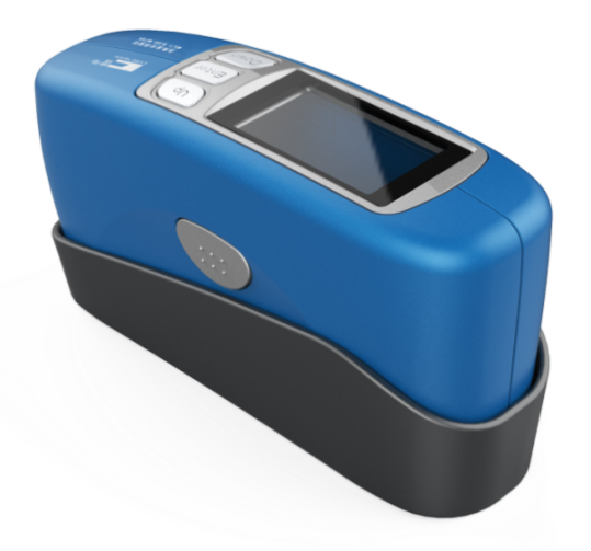
The Nanophotometer N50 allows the rapid assessment of DNA, RNA and protein concentrations of small (1 µl) samples. It is a stand-alone instrument for nucleic acid quantification and purity check (dsDNA, ssDNA, RNA, mRNA, miRNA, Oligos including dye-labeled samples)) and for protein quantitation and purity check (BSA, Antibodies, IgG, IgE, Serum Albumin, etc., including dye-labeled samples), small molecules in organic solvents, kinetics in a drop, and more. It has an interactive touch screen, and a built-in high performance computer processor and sufficient memory to provide a powerful and easy storage of methods and data.
Location: FA 314 (door code access required)
The Sartorius Octet R8 label-free protein analysis system provides fast, high throughput, and accurate characterization for biomolecules such as proteins, antibodies, peptides, DNA, RNA, liposomes, viruses and various media. It performs quantitation and kinetic analysis of up to 96 samples in 30 min to 2.5 hours, depending on the specific assay. It can be used for a wide range of analyses, including kinetic analysis, titers IgG and other proteins, reagent quantification, crude antibody screening, epitope binning/mapping, ligand binding assays, infectious disease monitoring, and more. Analysis can be done using a single channel or up to 8 channels, enabling flexibility in sample throughput.
Note: Please acknowledge the funding source NIH S10 OD032385 in publications and presentations when using this instrument. Users must purchase their own consumables
Location: FA 312 (door code access required)
The QIAgility is a compact benchtop instrument that enables rapid, high-precision setup of PCR experiments with ready-to-use software for ease and convenience. The software provides step-by-step guidance for worktable setup and automatic calculation of all mixes, eliminating the need to program pipetting steps. Just select the kit and cycler, define your targets and start the run. For added flexibility, the QIAgility is operated via a laptop computer.
Location: FA 314 (door code access required)
The Qubit 4 Fluorometer accurately measures DNA, RNA, and protein quantity. The Qubit together with an RNA IQ Assay Kit can accurately distinguish intact from degraded RNA in just two easy steps. For all Qubit assays, the concentration or quality of the target molecule in the sample is reported by a fluorescent dye that emits a signal only when bound to the target, which minimizes the effects of contaminants on the result. The easy-to-use touch-screen menus make it easy to select and run the assays you need, with results displayed in just a few seconds.
Location: FA 318 (door ‘key’ access and laser certification required)
Two-photon microscopy has become an indispensable tool for cellular neuroscience, allowing high-resolution fluorescent imaging of cells in vitro and in vivo living brains and whole animals. The HyperScope system is a custom-build, two-photon microscope equipped with a Mai-Tai eHPDS broadly tunable Ti-sapphire laser (690-1040 nm), multiple objectives (16x, 20x and 40x), and stands on a Thorlabs breadboard and anti-vibration table. With the Scientifica SciCam Pro CCD camera, it is possible to perform functional imaging of video frame rates of 24 fps (full resolution) and 40 fps (binned 2 x 2). The detector assembly includes one red-shifted PMT and one GaAsP PMT module, and epifluorescence imaging with filter sets for GFP and mCherry. The scan mirrors in the imaging path can be arranged in multiple configurations depending on your needs. With up to 3 mirrors in series, you can choose between galvo/galvo (GG), resonant/galvo (RG), or resonant/galvo/galvo (RGG) arrangements. The microscope is fitted with an ultra-smooth Z-axis drive to ensure vibration-free movement, with the Z-axis being controlled with software for acquiring z-stacks with a minimum step size of 0.1 mm. The addition of a Piezo z-axis objective positioner allows volume imaging up to 400 mm. The microscope can be easily configured and switched between in vitro and in vivo applications by removing the substage optics and riser block. The system is enclosed in a black-out cage to prevent light and noise pollution of biological samples.
This platform includes SciScan acquisition software, allowing users to integrate SciScan easily with custom-written applications, such as LabView and ScanImage. The ScanImage software interface used by Scientifica was developed by HHMI Janelia Farm specifically for neuroscience applications and is considered to be the most advanced software for two-photon laser scanning on the market.
Location: FA 312 (door code access required)
The GeneSys spectrophotometer performs high-throughput quantitative UV-Vis measurements where a double beam is required as a reference cell position. Optimized for usability and performance, this system features a high-resolution color touchscreen and optional Wi-Fi networking. The compartment is an 8-position cell changer (standard).
Location: FA 314 (door code access required)
The Trans-Blot Turbo transfer system combines traditional blotting techniques with modern filter paper and buffers allowing rapid transfer of proteins from gels to membranes with minimal preparation time. The system integrates a high amperage power supply that directs current between a built-in platinized titanium anode and a stainless steel cathode.
Location: FA 314 (door code access required)
The NanoCellect WOLF G2 cell sorter is used for a variety of applications, including antibody discovery, single cell sorting and plating, genomics, single-cell RNA sequencing, plant biology, gene editing, cell line development, and discovering microorganisms in ocean or pond water. The WOLF G2 is designed to gently sort viable, healthy cells using a sterile cartridge-based microfluidics system. The WOLF G2 cell sorter is ideal for tasks like cell line development and sorting plant protoplasts, as well as for monoclonal outgrowth of iPSCs and microalgae. At < 2 psi, the WOLF G2 cell sorter is gentler than any conventional cell sorters, enabling healthier cells post-sort, especially for engineered lines, primary cells, and stem cells. All laser configurations afford <250 MESF sensitivity, along with forward and backscatter, providing as low as 1 μm resolution.
The ease of use and operation of the Wolf G2 utilizes disposable, aerosol-free microfluidic cartridges that allow for sterile sorting that protects the sample from the environment and scientists from the sample. The cartridges employ a piezoelectric actuator and operate at less than 2 psi allowing for gentle sorting that preserves viability and RNA integrity. Cartridges have a 100% disposable fluidic pathway, making it ideal for labs with many samples where cross-contamination or biohazard cleanup is an issue. The microfluidic cartridges are sterile and produce no aerosols, making it some of the safest sorting on the market. The cartridges also allow for maximum flexibility by allowing users to sort using their own buffers and media and only consume about 50 mL of sheath fluid in 1 day.
Note: users must purchase their own consumables, i.e. microfluidic sorting cartridges, calibration beads
For iLab onboarding, training of new users, and assisted use, please contact Andrew Yanez at ayanez@unr.edu. For general information about the core and support letters for grant proposals, please contact Dr. Alexander van der Linden (avanderlinden@unr.edu).

Life Science Equipment 1664 N. Virginia Street, Reno, NV 89557 Effie Mona Mack (EMM), Mailstop: 0296 kumataka@unr.edu (775) 784-4780